
Nearly silent MRI tracks mice reaching for food
SORDINO reduces noise during image acquisition and records brain signals in awake animals whose heads are fixed but whose paws remain free.
Leitura autorizada · 3 crédito(s) restante(s)
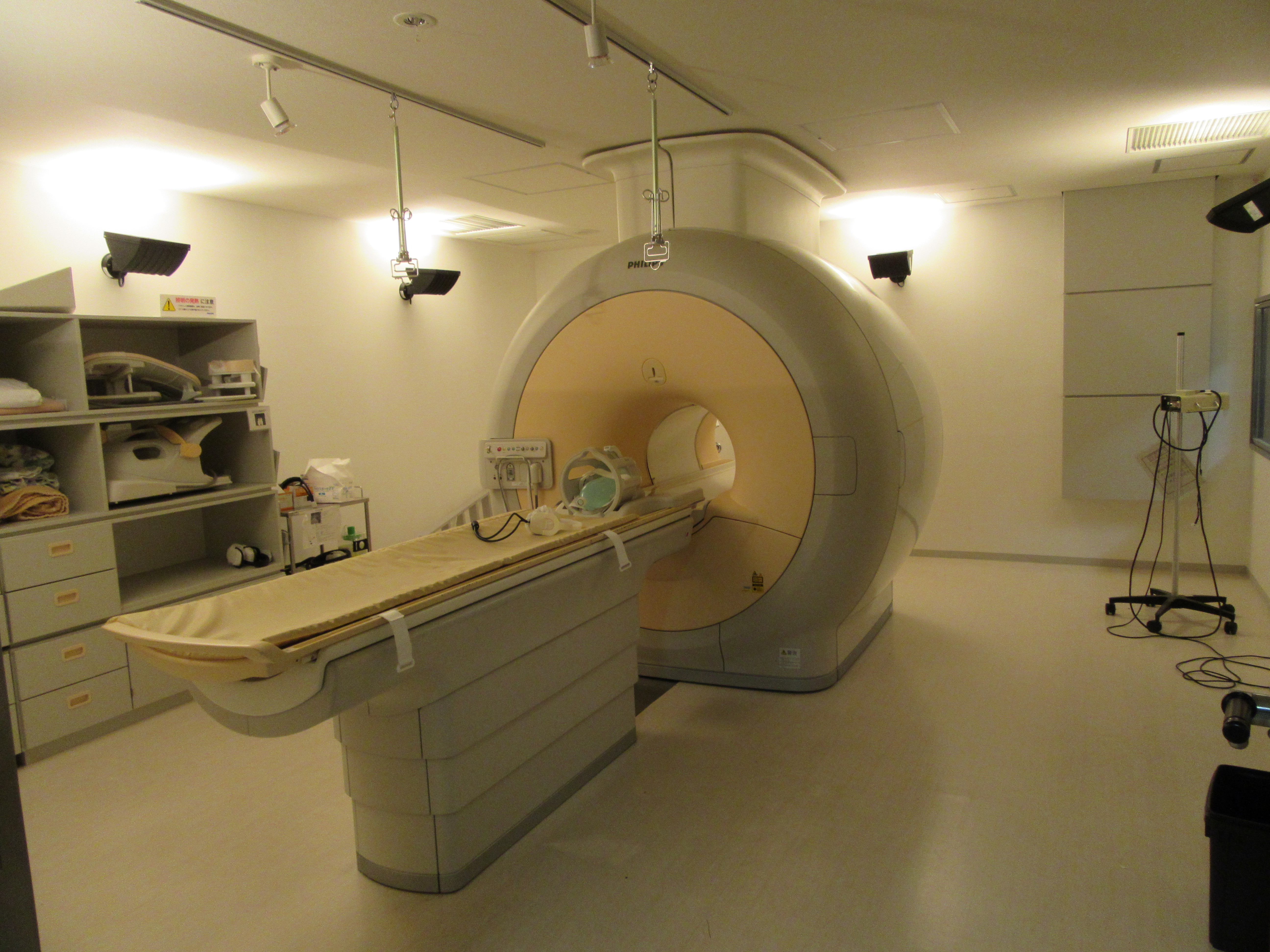
MRI noise and movement-induced distortions make it difficult to follow an animal’s brain in action. A team led by Yen-Yu Ian Shih at the University of North Carolina at Chapel Hill redesigned functional magnetic resonance imaging acquisition to address both obstacles. The technique, called SORDINO, was introduced September 9 in Nature Neuroscience and tested on an experimental 9.4-tesla scanner; teslas measure magnetic field strength.
To locate the origin of each signal, the scanner imposes small variations in the magnetic field across space. Abrupt changes in those variations make the coils vibrate and generate noise. SORDINO keeps their combined strength nearly constant and rotates their direction smoothly and continuously, collecting data throughout the change. The team compared the method with conventional acquisition, known as GRE-EPI, and with ZTE, an alternative that starts reading the signal almost immediately.
Using a microphone about three meters from the scanner’s center, the team measured how much each acquisition increased sound pressure levels relative to the idle scanner. Within the mouse hearing range, the average difference was approximately 0.1 decibel for SORDINO, 0.9 for ZTE, and 13.0 for the conventional technique, across 1,210 recording segments per condition. The cooling system continues to operate: the improvement is the near-elimination of additional acquisition noise, not absolute silence.
To move on to observing awake animals, the researchers built a small plate that fixes the head and also acts as an antenna to transmit and receive MRI signals. The body and paws remain free. After five days of habituation, 25 mice were each scanned for 30 minutes, with a three-dimensional image every two seconds and volume elements measuring 0.4 millimeters on each side. Analysis of fluctuations shared across regions recovered known patterns of the brain’s functional organization.
In a separate experiment, four mice learned to extend a paw and grasp a small piece of food presented every 20 seconds. An air-powered device placed the food within the animal’s reach inside the scanner. The analysis combined 24 sessions, with 75 to 100 grasping movements per session, and compared signals during and after the action with the seconds before food presentation. The head remained fixed throughout the task.
The maps showed an initial signal increase in the motor cortex opposite the paw being used, alongside a decrease in the retrosplenial area, located in the posterior, medial portion of the cortex. Over the following seconds, increases appeared in sensory and motor regions of both hemispheres, the thalamus, and the cerebellum, before returning to baseline. MRI follows changes in blood and oxygenation associated with neuronal activity. This image sequence is therefore not a movie of electrical impulses passing from cell to cell.
The advance combines a quieter acquisition environment with the ability to follow multiple brain regions during a coordinated action. For behavioral researchers, it expands the range of tasks that can be studied without anesthesia while covering the whole brain. The next challenge is to explore that range and establish how well accuracy and sensitivity hold up on other equipment and under other conditions.
Key points
- SORDINO smooths the changes used to locate signals and reduces acquisition noise.
- Four mice, studied across 24 sessions, enabled brain-signal mapping during a food-grasping task.
- The technique measures responses linked to blood and oxygenation; its use in humans has not yet been demonstrated.
Comments
No comments have been published yet.
Sign in with a subscription to comment.