
A small human circuit reproduces the waves of anesthesia
Three-dimensional models of nerve cells responded to propofol with quieter neurons and coordinated slow waves. The result helps probe the mechanism without measuring consciousness.
Leitura autorizada · 3 crédito(s) restante(s)
General anesthesia makes surgery possible, but connecting a drug's action on individual cells to the electrical waves seen in an anesthetized brain remains difficult. Daniel Toker, Ranmal Samarasinghe and colleagues at the University of California, Los Angeles (UCLA), found that connection in a human model grown in the laboratory: after exposure to propofol, an anesthetic drug, individual nerve cells fired less often while activity across the circuit began to oscillate in slow, synchronized waves. The study was published in the British Journal of Anaesthesia; Tiare Dunlap reported the results for UCLA Newsroom on October 1.
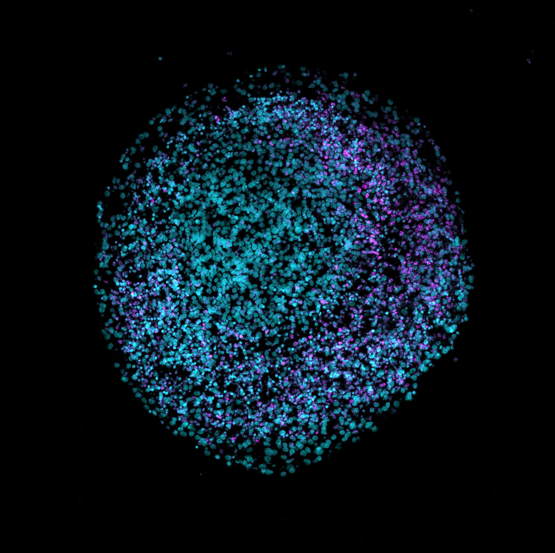
In simple terms, the team built small three-dimensional circuits called assembloids: combinations of tissues derived from adult cells reprogrammed to return to a stem-cell-like state. They joined models of regions that produce excitatory neurons, which propagate signals, and inhibitory neurons, which dampen signals, with supporting glial cells. These circuits make it possible to observe the activity of individual cells and the rhythm of the group at the same time. They are simplified models of developing tissue, not complete brains.
The team exposed these circuits to propofol, a drug used to induce general anesthesia, and recorded their electrical activity. Although each neuron became less active, many began to fluctuate together, producing broad, slow waves similar to those seen in electrical recordings from anesthetized patients. Coordination, rather than increased firing by individual cells, explains how the collective signal can grow as the cells become quieter.
Two controls help locate the mechanism. Simpler models without inhibitory neurons did not generate the characteristic pattern. When the researchers blocked the cellular receptors on which propofol acts, the response also disappeared. Because these assembloids have no thalamus, a deep brain structure that exchanges signals with the cerebral cortex, the brain's outer layer, the experiment shows that a minimal cortical circuit can be sufficient to produce this electrical pattern in the laboratory. It does not show that the thalamus plays no part in anesthesia in people.
The result is a controllable platform for asking which cells and signals generate rhythms associated with anesthesia. The evidence supports the reproduction of an electrical signature and the role of inhibitory neurons in this model; it does not prove that the tissue is conscious or, by itself, explain the loss of human consciousness. Comparisons across anesthetics and patient recordings will be decisive in establishing how far this mechanism generalizes.
Key points
- Propofol reduced the activity of individual neurons and synchronized slow waves in the cultivated circuit.
- Without inhibitory neurons, or when propofol receptors were blocked, the pattern did not appear.
- The model reproduces an electrical signature, not consciousness or the entire adult brain.
Comments
No comments have been published yet.
Sign in with a subscription to comment.